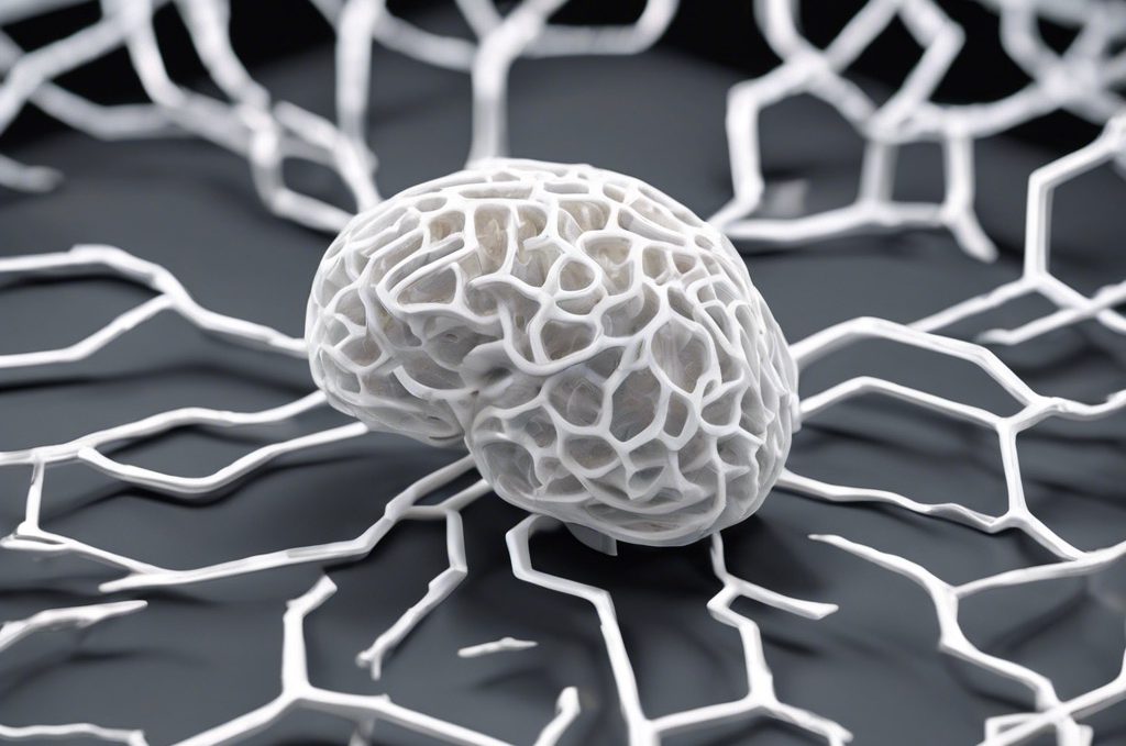
Towards Printing the Brain
“The high resolution of two-photon polymerisation makes it possible to print details in the micro- and nanometre range and is therefore very suitable for imaging cranial nerves.”
Medical University of Vienna
Magnetic resonance imaging (MRI) has become an indispensable tool for examining the living human brain in both research and clinical settings. However, one limitation has been difficulty rigorously validating MRI findings against known gold standards. Now, a team of scientists in Europe has demonstrated a breakthrough solution—using high-precision 3D printing to fabricate biomimetic “brain phantoms” that can serve as ground truth models for validating MRI techniques like diffusion MRI (dMRI). Their work, published in Advanced Materials Technologies, shows significant potential for advancing MRI as a non-invasive probe of brain structure and connectivity.
Validating neuroimaging results is crucial but challenging, as there are obvious ethical concerns with conducting truly “gold standard” comparisons on the same brain tissue sample using multiple methods, some of which are invasive. To address this, researchers design simplified artificial test objects known as “phantoms” that mimic tissue properties. However, conventional phantom fabrication methods were unable to achieve the combinations of small feature sizes, tight control over microstructure, and large sample dimensions needed for experiments in human MRI scanners.
Enter two-photon polymerization (2PP) 3D printing. Considered the state-of-the-art in micro- and nanoscale additive manufacturing, 2PP achieves extraordinarily high resolution by using ultrafast laser pulses to sculpt solid objects just a few microns across, layer by layer, from liquid resin. However, structures fabricated with conventional 2PP have been limited to just a few hundred micrometers in size.
The research team at the Medical University of Vienna and TU Wien overcame this limitation through multiple strategies. First, they mounted the photoresin directly on the 3D printer’s focusing objective and lowered it incrementally, allowing structures to be built taller. Second, they printed in successive “tiling areas” to stitch together building blocks spanning larger xy areas. By combining these techniques, they achieved phantoms with dimensions on the millimeter scale—big enough for a human MRI—while retaining microscale features as small as 12 μm across, comparable to axon diameters in white matter.
Using their optimized 2PP process, the team fabricated two proof-of-concept phantoms. The “sandwich” phantom contained parallel channels in three alternating orientation layers, while the “wafer” phantom had rows of channels alternating between two perpendicular directions within each voxel volume. Both phantoms incorporated over 14,000 individual channels each to mimic human white matter density and provide adequate signal for MRI.
Imaging and analysis showed the 2PP phantoms were compatible with MRI and provided appropriately anisotropic diffusion signatures. Within each phantom, voxel-wise ball-and-stick modeling correctly identified the predetermined channel orientations. When processed with fiber tracking algorithms, the results agreed with the designed parallel and crossing arrangements. Measurements validated that the materials induced negligible magnetic field distortions.
Beyond serving as a demonstration of 2PP printing capabilities, these initial phantoms open doors for improving dMRI validation. Future work could incorporate curving or kissing fiber geometries that better approximate human white matter complexity. Each customized phantom would then act as ground truth against which MRI findings could be benchmarked. This level of tight control over microstructural parameters has never before been possible in dMRI phantoms.
According to the researchers, their results establish new standards for the construction of 3D biomimetic scaffolds. The versatility of 2PP fabrication allows essentially any arbitrary axon tract geometry to be replicated with precision nearing that of real neural tissues. With further optimization, even larger and more anatomically accurate brain phantoms may be achievable.
By enabling accurate dMRI ground truth modeling, such phantoms have far-reaching implications. They will enhance efforts to determine sensitivity and specificity for detecting disease states or differences based on white matter microstructure. Importantly, they could validate specialized analysis techniques like white matter tractography mapping, which is challenging today without controlled test cases. This would strengthen arguments for clinical applications and drive more reproducible neuroscience research. Ultimately, the new brain phantoms herald tangible improvements to noninvasively probing human brain connectivity using MRI.
Through creative application of sophisticated 3D printing, this research demonstrates a major technical achievement and provides a framework for addressing key validation needs in the field of diffusion MRI. By fabricating accurate biomimetic tissue models with unprecedented control over microstructural properties, it establishes a critical foundation that should supercharge progress in both developing and applying this powerful neuroimaging method. Now that they have shown the feasibility and utility of high-resolution brain phantoms produced by two-photon polymerization 3D printing, the researchers plan to refine the approach so its full potentials can be realized. Their work exemplifies how technologies once considered too limited can be ingeniously adapted to open up new scientific possibilities.
Reference(s)
- Michael Woletz, Franziska Chalupa‐Gantner, Benedikt Hager, Alexander Ricke, Siawoosh Mohammadi, Stefan Binder, Stefan Baudis, Aleksandr Ovsianikov, Christian Windischberger, Zoltan Nagy. Toward Printing the Brain: A Microstructural Ground Truth Phantom for MRI. Advanced Materials Technologies, 2024; 9 (3) DOI: 10.1002/admt.202300176
Click TAGS to see related articles :
3D PRINTING | BRAIN | DIAGNOSTICS | NEUROLOGY
- Millions of new solar system objects to be found...on June, 2025 at 1:34 am
Astronomers have revealed new research showing that millions of new solar system objects are likely to be detected by a brand-new facility, which is expected to come online later this year.
- Tea, berries, dark chocolate and apples could...on June, 2025 at 3:50 pm
New research has found that those who consume a diverse range of foods rich in flavonoids, such as tea, berries, dark chocolate, and apples, could lower their risk of developing serious health conditions and have the potential to live longer.
- Being in nature can help people with chronic back...on June, 2025 at 3:50 pm
Researchers asked patients, some of whom had experienced lower back pain for up to 40 years, if being in nature helped them coped better with their lower back pain. They found that people able to spend time in their own gardens saw some health and wellbeing benefits. However, those able to immerse […]
- Scientists say next few years vital to securing...on June, 2025 at 3:50 pm
Collapse of the West Antarctic Ice Sheet could be triggered with very little ocean warming above present-day, leading to a devastating four meters of global sea level rise to play out over hundreds of years according to a new study. However, the authors emphasize that immediate actions to reduce […]
- First direct observation of the trapped waves...on June, 2025 at 3:50 pm
A new study has finally confirmed the theory that the cause of extraordinary global tremors in September -- October 2023 was indeed two mega tsunamis in Greenland that became trapped standing waves. Using a brand-new type of satellite altimetry, the researchers provide the first observations to […]


